Benign vs. Malignant Tumors
- benign : 팽창(expansile), 주변 조직을 누름, 명확하게 구분됨
- malignant : 전이(invasive), 명확하지 않음(diffuse)
- 계속 복제됨 (unlimited replicative potential)
- exogenous 성장 자극 인자와는 별도로 성장, 성장 억제 신호에는 insensitive
- benign cell에 비해 apoptotic cell death도 덜함
- marked angiogenesis를 자극해 혈관 전이를 촉진
| 특성 | Benign | Malignant |
|---|---|---|
| Differentiation | well-differentiated morphologic (명확) | poorly differentiated (명확하지 않음, diffuse) |
| origin과 비슷한 조직 구조 | 때로 origin이 불명확 | |
| anaplasia(퇴화) 거의 없음 | 다양한 단계의 anaplasia | |
| Growth rate | 느리고 점진적 | 빠른 성장 |
| mitotic figure | 드물고, 정상 | 빈번하고, 비정상 |
| necrosis | 거의 없음 | 혈액 공급 저하 시 발생 |
| Local invasion | 있는 자리에서 응집, 팽창성 | 주변으로 전이 |
| capsule 있을 수 있음 (경계 명확) | capsule 없거나 불명확 | |
| Metastasis | 없음 | 종종 있음 |
Differentiation
(tumor 생길 시 형태가 어떻게 변하는가)
Morphology
-
잘 구별된 원래의 모양을 잃고 흐트러진 모양 , 정상 세포에 비해 morphologic variability가 큼
-
매우 큰 nuclei(karyomegaly)가 관찰되거나, nuclear size 다양(anisokaryosis), shape와 chromatin 분포 패턴이 다양하거나, multiple nuclei를 가지기도 함
-
빠른 세포 성장과 분열을 하려면 ribosome이 많이 필요하기 때문에 호염기성을 띰(basophilic cytoplasm)
-
정상 구조를 점점 잃음, continual renewal을 거치다 보면 normal maturation sequence가 변형되기도 함
-
악성 종양은 양성보다 덜 구별됨(less differentiated)
-
특히 악성 종양에서 anaplasia(역분화), atypia(이형성) 나타남
- anaplastic cell : poorly differentiated, cell size 다양(anisocytosis), cell shape 다양(pleomorphism)
- anaplastic nuclei : 잦은 분화로 염색체가 응축되어 darkly staining(hyperchromatic), 세포질에 비해 핵이 유난히 큼
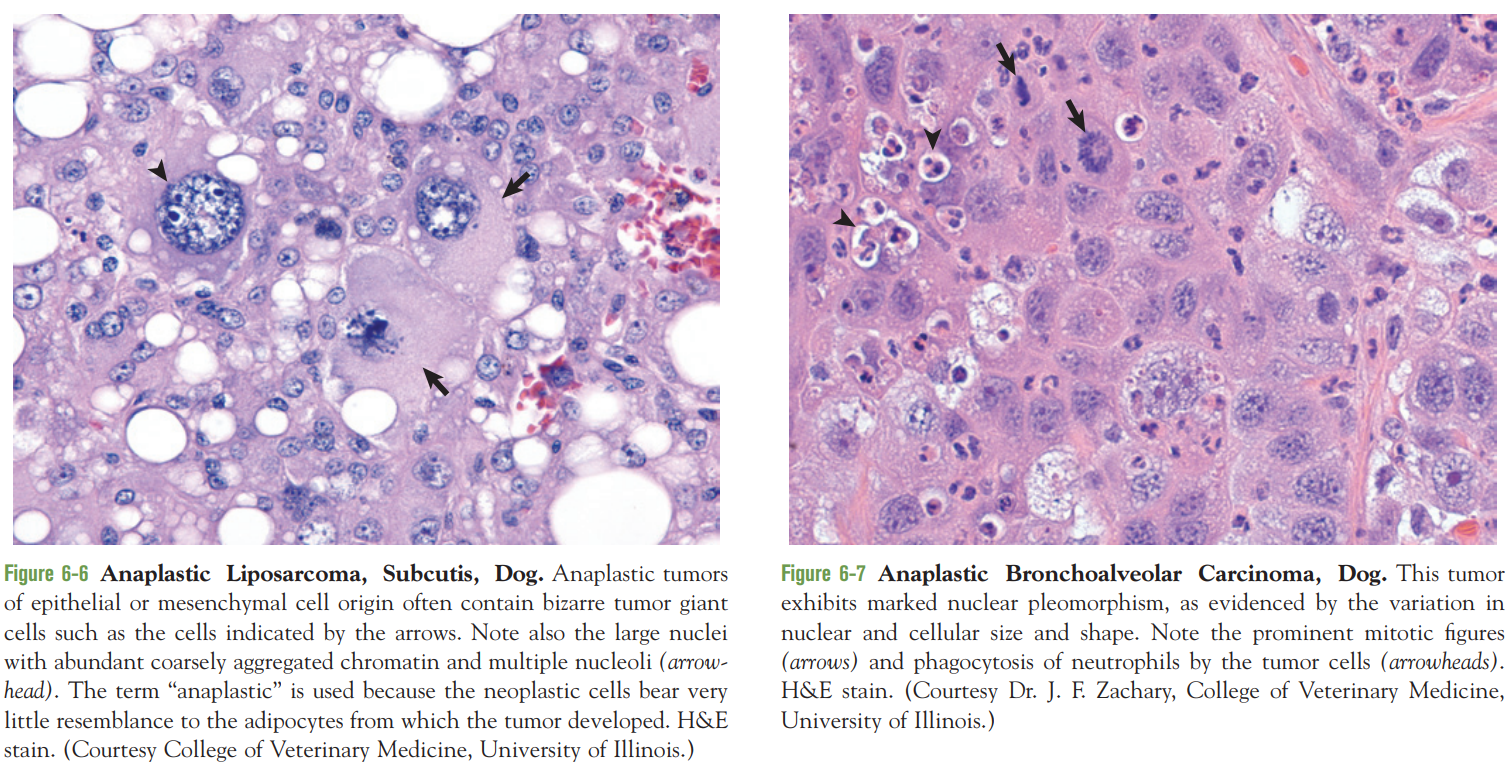 (왼) 화살표 : giant cell, 화살표 head : 엄청 큰 핵 / (오) 화살표 : mitotic figures(giant)
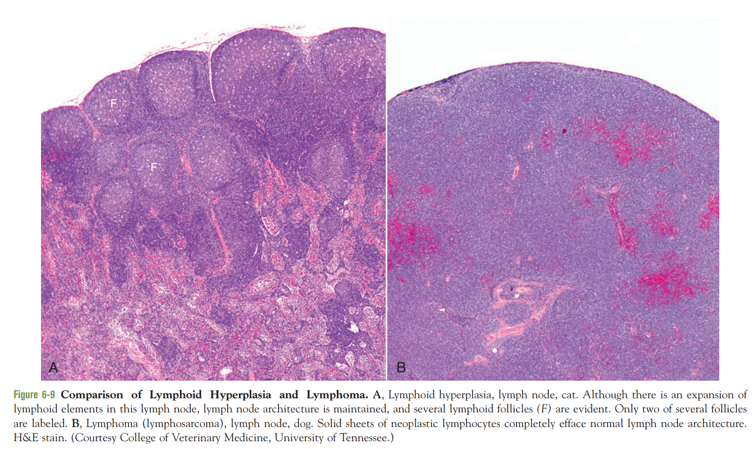 (왼) Hyperplasia / (오) Sarcoma
Function
- 기능의 loss는 모양(differentiation)의 loss와 자주 동반됨
- ex) 소장 상피의 neoplastic cells는 microvilli가 없다 → 흡수 능력도 없음
- 종종 normal function이 유지되기도 함
- ex) thyroid adenoma가 계속 갑상샘 호르몬 생성, plasma cell tumor가 immunoglobulin 분비
- 보통 정상적인 regulatory pathway에 대한 반응성을 잃어 적절히 기능하지 못함
Proliferation
Tumor growth
- unlimited proliferative potential
- 많은 종양세포는 immortal
- 외부 성장 자극/억제 인자의 영향X, apoptotic signal을 듣지X → cell division의 한계 넘어 분열
Cell division
- cell cycle은 G1, S, G2, M^[각각 G0: presynthetic, S: synthetic, G2: premitotic, M: mitotic] phase로 구성.
- Quiescent cells는 G0에 머물면서 cell cycle에 들어가지 않음 (외부 인자의 자극 없는 이상)
- DNA damage가 발생하면, 정상적으로 분열하는 세포들도 cell cycle arrest를 거침 (cell cycle checkpoints들이 있음) → cell cycle arrest는 p53^[multifunctional tumor suppressor gene product]에 의해 개시되며, 세포에게 DNA damage를 복구할 시간을 줌
- neoplastic cells은 G0으로 보내는 신호에 반응X, p53 발현X → 계속 cell cycle → DNA damage 이후 cell cycle arrest를 겪지 않으므로, 점점 mutagenic DNA damage가 축적됨
- mitotic index
- 응축된 염색체와 핵막 소실이 관찰
- 세포분열이 일어나고 있다는 지표로, 종양의 malignant potential을 나타냄
- 세포분열을 멈출 수 없는 cancer cells에서 mitotic figures가 나타남 (abnormal figures도 관찰)
- 정상 세포는 끊임없이 주변 환경과 대화하며 항상성을 유지하는 반면, neoplastic cells는 성장 자극 물질도, 성장 억제 신호도 받지 X → 주변 환경에 필요한 것을 제공해주지 않고, 계속 복제만 함
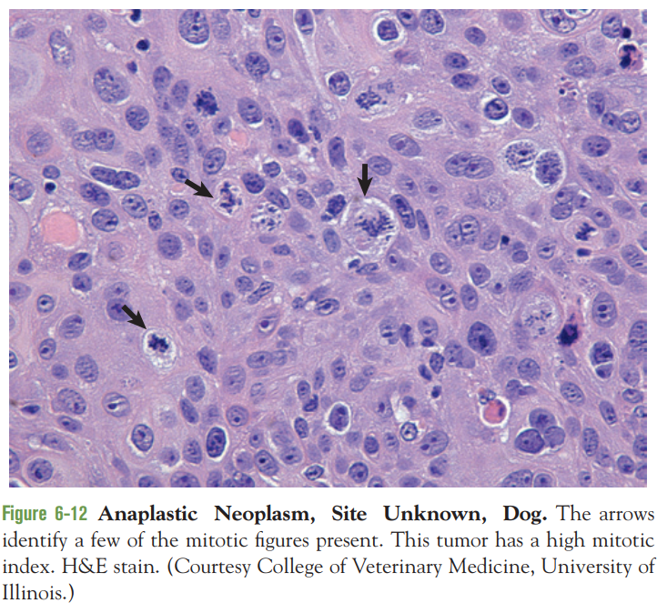 화살표: mitotic figures / high mitotic index를 가짐
Cell death
Senescence(노화)
- DNA damage, oxidative stress, telomere shortening 등에 의해 나타남. (신호: p53 활성화, retinoblastoma pathway)
- DNA template의 말단에 있는 telomere는 복제되지 않기 때문에, 세포 분열 때마다 짧아짐. telomere가 극히 짧아지면 더 이상 분열할 수 없음(⇒senescence) ^[예외: telomerase를 발현하는 embryonic cell 때만 telomere가 계속 복제됨]
- neoplastic cells는 telomerase를 생산할 수 있음 → 세포 노화를 겪지 않고 immortality 획득
Apoptosis(세포자살) (“programmed cell death”)
- hypoxia, essential nutrients의 부족은 apoptosis로 이어질 수 있음. DNA damage가 발생하면 p53에 의해 apoptosis 야기. cytotoxic immune cells(T림프구, NK cell)가 apoptosis를 자극
- 염색체의 margination, 핵의 condensation과 fragmentation, organelles를 보존하는 cell condensation 등은 apoptosis의 morphologic hallmarks가 됨
- 결국 세포는 membrane-bound apoptotic bodies로 쪼개져서 surrounding cells에게 잡아먹힘. 이때 염증 반응은 자극하지 않음.
- 정상적으로 모든 세포는 apoptosis를 겪을 수 있지만, 암세포는 저항성이 있음.
- p53 gene inactivation, survival signal 활성화, death factor 신호전달 억제를 통해 apoptosis를 우회함
Autophagy(자가포식)
- degeneration of a cell’s own organelles within autophagosomes
- 영양소 결핍 시 cell survival의 기전으로 사용될 수 있으나, 광범위한 autophagy는 apoptosis 유도
- 보통 tumor cell에서 autophagy는 억제되어 있음
- mTOR inhibitor는 autophagy를 증가시켜 암 치료제로서의 가능성을 가짐.
- mammalian target of rapamycin(mTOR) kinase : autophagy를 억제